ORINS Standard Scanning Manikin (mid 1950s)
This scanning phantom/manikin was developed by Marshall Brucer at the Oak Ridge Institute for Nuclear Studies (ORINS) sometime in the mid 1950s. This particular example, the second in what became a family of twenty (or possibly more), was given the name Bonnie Boleyn. As you might guess, Anne Boleyn was the first. In 1957 there were only these two, Anne and Bonnie, but later on, Chloe, Drusilla, Eupexia, Felicia, Grenadine, Hortense, Ibis, Jezebel, Katrinka, Lulu, Moira, Nabby, Ophelia, Pandora, Queenie, Rhoda, Sara Coma, Terry Toma were added.
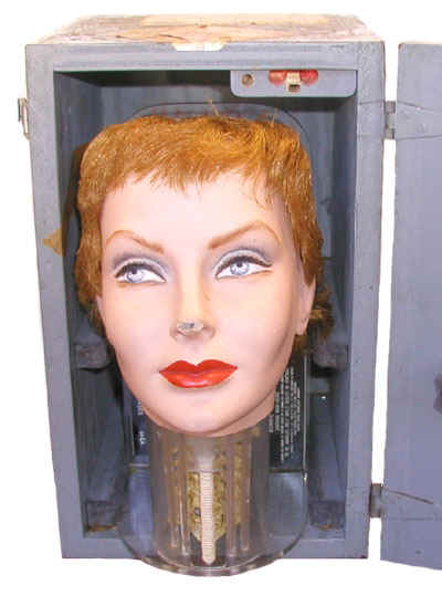
Brucer described the first scanning manikin, Anne Boleyn, as follows: "the Anne Boleyn manikin consists of a head and neck. There is nothing in the head. At the base of the head is an artificial neck. Contained within the neck is a movable mock thyroid gland, two fixed metastases, a mock spinal cord filled with mock-iodine... and two low-level sets of simulated blood vessels. In the thyroid is a cold spot simulating a nonfunctioning adenoma of the thyroid gland."
Bonnie Boleyn (photo to right) contained 10 uCi in the thyroid, 2 uCi in each of the blood vessels, 5 uCi in the spinal column and 1 uCi in each of the two metastases.
These manikins were shipped to medical facilities around the world to assist in establishing the optimal operating parameters of rectilinear scanners, and to assist personnel in the interpretation of the scan.
A rectilinear scanner (see photo left) consisted of a collimated NaI detector that moves back and forth over the body of a patient who has been administered a radiopharmaceutical. The output was used to generate an image on a piece of paper or photographic film.
The quality of the image, which impacted the physician's ability to interpret the scan, depended on, among other things, the scan rate, the size of the dots used to produce the image, the type of collimator and the administered activity.
Regarding the need for such a phantom, Marshall Brucer wrote: "Theoretical physicists are a notably visionary lot and electronic physicists are in league with the devil. Their importance to medical instrumentation has been proved with nauseating repetition and needs no further comment. However, they have never learned the necessity for controlled experiments. The general practice up to now in comparing which scans were good and which scans were bad has been to take a number of scans on a number of patients and to discourse on the prettiness of the picture. Since one never knows what the patient really had, the voice of authority is not the patient but the loudness with which the physician talks."
Brucer's family of manikins provided a means by which a physician's skills could be put to the test.
References
- Marshall Brucer. Medical Instrumentation in Radiation Biology and Medicine. edited by W.D. Claus. Addison-Wesley Publishing Company. Reading, Mass. 1958.
- Marshall Brucer. In Search for the Hole. Unpublished.
- Marshal Brucer. Radioiodine Uptake Measurement. Apartado del Acta Radiologica Interamericana Vol. VII, No. 3-4: 129-143. 1957.
- Oak Ridge Institute of Nuclear Studies. Medical Division Midyear Report for the Period Ending December 31, 1956.

ORAU historical spotlight: The story of Marshall Brucer, a physician and nuclear medicine pioneer
Learn more about William Brucer M.D., who was one of the early figures in the history of ORINS, which later became ORAU.