Model of ORINS Fixed Geometry Training Table (1955)
This wood and masonite model was built in 1955 for Marshall Brucer, the then head of the Medical Division at the Oak Ridge Institute for Nuclear Studies (ORINS). It was one of several such models showing the various types of equipment in use at ORINS. These models were used in traveling exhibits and lectures as a way to convey a three-dimensional idea as to what the equipment was like.
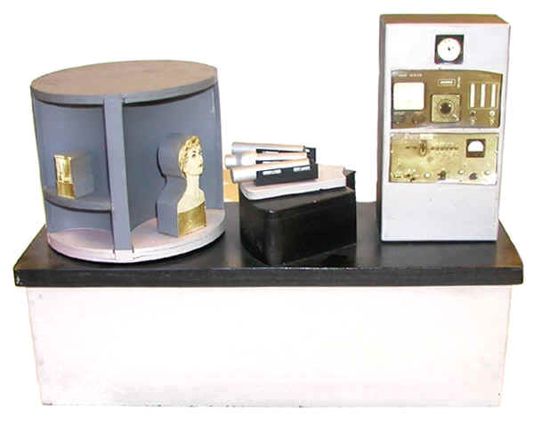
This particular model shows an instrument for measuring the activity of either iodine or mock iodine in the neck, head, and torso phantoms that ORINS distributed to medical facilities as a means to calibrate their detector systems. The model indicates that three different detectors (the horizontal silver cylinders in the center of the model) were used for this purpose. The detector in the center with the enlarged end (a collimator) would have been a sodium iodide detector. The other two would have been GM detectors. Most likely one was an end window GM and the other was a bismuth side window GM. The detector output was recorded by a Tracerlab 1000-decade scaler (immediately below the timer on the right side of the model) and an Instrument Development Laboratory Model 161 binary scaler. An IDL Model 162 binary scaler can be seen in the collection.
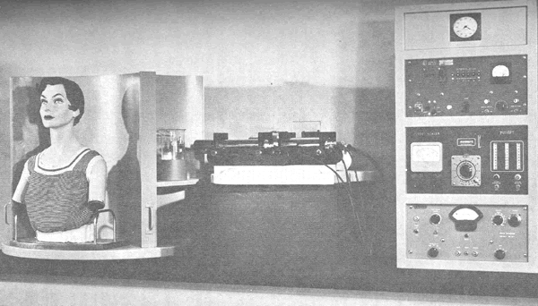
Quoting Marshall Brucer et al (1956): "to facilitate the study of these mannequins and to demonstrate the different results that may be obtained in thyroid uptake measurements by the use of various instruments, a fixed-geometry training table has been set up... The use of this fixed-geometry training table is open to professional visitors to the Medical Division of ORINS." The mannequins being referred to were like that shown in the above photo. Mock iodine was incorporated into the mannequins' "thyroids" and the mannequins were then shipped to hospitals around the world to assist in the calibration of the instrumentation being used for thyroid uptake measurements.
Since the instrumentation at the various hospitals employed different types of detectors (e.g., NaI, end window GMs and side window GMs), it was necessary to know if the detector type affected how well mock-iodine, a mix of Ba-133 and Cs-137, served as a substitute for real iodine-131. Hence the use of different detector types in the training table.
The builders of the model, Dewey Ferguson and Harry Kimble, added their signatures and the date (1955) to the left end of the model.
Size: 16" long, 6 1/2" wide, 12 1/2" high
Donated by Harold Hodges.
References
- Marshall Brucer, T.H. Oddie and James S. Eldridge. Thyroid Uptake Calibration. I. Mock-Iodine, A Radioactive Iodine Gamma-ray Standard. ORINS-14. July 25, 1956.
- Elizabeth B. Anderson. Ten Years With the Peaceful Atom. September 1946 to September 1956. Unpublished presentation to the Parent Teachers Association, Park Ridge, New Jersey, October 9, 1956.