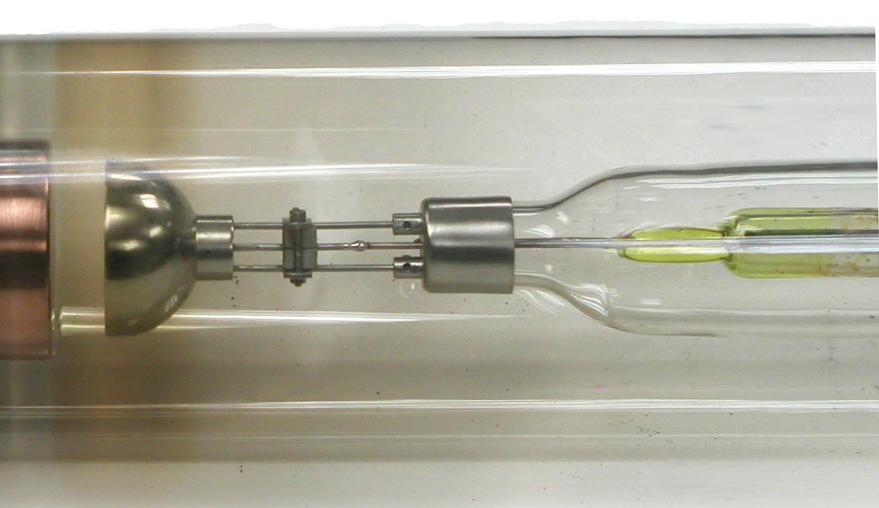
General Electric CA-2 X-ray Diffraction Tube (ca. 1930s)

This is a General Electric Model CA-2 tube designed for x-ray diffraction analysis. I believe that the "CA" stood for crystal or crystalline analysis. The "2" indicated that this was a fine to medium focus tube.
For X-ray diffraction analysis, the X-ray beam needed to be nearly monoenergetic. This was accomplished by using the target's characteristic X-rays and selectively filtering out the bremsstrahlung. The most common target material was molybdenum which would emit characteristic X-rays near 18 keV.
It would typically have been operated at a potential of 30 kVp with a 20 mA current. The maximum rating for continuous operation was 42 kVp with a 25 mA current.
The two steel tubes seen on the left end of the tube in the top photograph would be connected to the cooling water supply.

In a tube designed for medical applications, the target would be sloped so that the X-rays would leave one side of the tube in a conical beam. In this tube however, the molybdenum target (not visible but embedded in the copper block on the left side of the photo) is positioned at right angles to the electron beam. The result: a uniform emission of x-rays in all directions in the flat plane of the target. That is why the cathode filament is a flat spiral similar to that in GE's "Universal" tube, rather than the Benson-style cylindrical coil. The brownish ring formed in the glass envelope indicates the path by which the X-rays exited the tube.
The cathode (not visible) is located in the hemispherical focusing cup just to the right of the target on the left side of the photo.
Notice that the glass stem holding the wire leads for the cathode (on right side of photo) is made of yellow uranium glass.
Size: Approximately 19" long, 2.5" diameter (max)
Kindly donated by Margie Wheaton in memory of her father Dr. Julius E. Marfy.
Reference
General Electric X-ray Corporation. Coolidge X-ray Tubes - Kenotrons. Bulletin No. 293. 1934.
