Universal X-Ray Tubes
The Universal Tube was so named because it could be used in almost any application, e.g., diagnostic X-rays (roentenography), fluoroscopy and therapy as well as various non-medical uses. In essence, it is the original Coolidge tube with a few minor modifications.
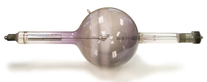
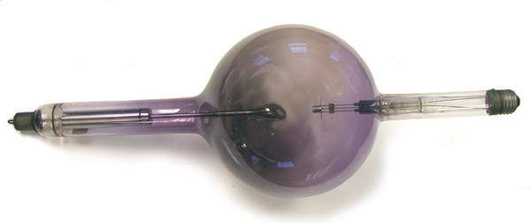
During operation, the heat from the anode is radiated out through the bulb of the tube. So that the glass won’t overheat, the bulb has to be kept fairly large: ca. 6-7” in diameter.
The threaded screw-in connector at the end of the cathode arm is the same as that employed with a light bulb. This reflects the fact that William Coolidge’s inspiration for the hot cathode X-ray tube was his earlier work developing tungsten filaments for incandescent lights.
The anode is solid tungsten and it is attached to a molybdenum stem. The latter, in turn, is connected to a split metal tube that helps provide support.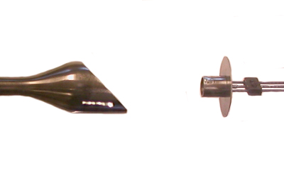
There were three variations on the Universal tube: those with a fine focal spot, those with a medium focal spot, and those with a broad focal spot. The cathode filament of the fine focus version forms a flat spiral near the end of a short molybdenum tube, while that of the medium and broad focal spot tubes forms a slightly conical spiral.
The following table indicates the ratings for these tubes.
| Tube Type | Maximum Voltage | Equivalent Spark Gap | Maximum Current | Continuous use limit |
|---|---|---|---|---|
|
Fine Focus |
100,000 |
6” |
25 mA |
20 seconds |
|
Medium Focus |
100,000 |
6” |
40 mA |
15 seconds |
|
Broad Focus |
100,000 |
6” |
80 mA |
5 seconds |
|
Broad Focus |
140,000 |
10” |
5 mA |
continuous |
The fine and medium focus tubes were better for general radiographic work. The broad focus Universal tube was best suited for therapeutic use because of its greater potential output. When the broad focus tube was operated at very high voltages, it would have been particularly important to keep the tube clean and far enough away from metal objects to prevent sparking.
The Universal tube needed to be operated with a rectified current. If unrectified AC current were used, electron emission from the tungsten target would be possible during the half of the cycle when the target ("anode") had a negative charge and the “cathode” a positive charge. This could only occur if the target had been heated to the point that it emitted electrons.
References
- E. C. Jerman. Modern X-Ray technic. Bruce Publishing Co. St.Paul. 1928.
- Radiology – An Illustrated History.
- W.D. Coolidge. The development of modern roentgen-ray generating apparatus. Am. J. Roentgenology. 24: 605-620. 1930.
- M.J. Gross, Z.J. Atlee. Progress in the design and manufacture of x-ray tubes. Radiology:365-377; 1933.
-
General Electric Universal X-ray Tube General Electric Universal X-ray Tube
-
Victor X-Ray Corp. Universal X-ray Tube Victor X-Ray Corp. Universal X-ray Tube